 Ciro Santilli
It is quite cool that photosynthesis works just like cellular respiration by producing a proton potential through chemiosmosis.
It is important to note that due to horizontal gene transfer, the early days of life, and still bacteria to this day due to bacterial conjugation, are actually a graph and not a tree, see also: Figure "Graph of life".
Definitely have a look at: coral of life representations.
TODO vs Phylogenetic tree? www.visiblebody.com/blog/phylogenetic-trees-cladograms-and-how-to-read-them:
Cladograms and phylogenetic trees are functionally very similar, but they show different things. Cladograms do not indicate time or the amount of difference between groups, whereas phylogenetic trees often indicate time spans between branching points.

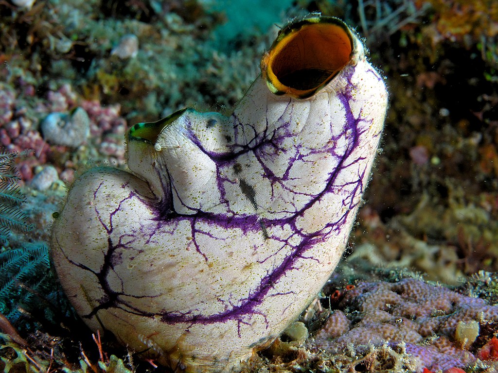
Coral of life by János Podani (2019)
Source. Fantastic work!!! Some cool things we can easily see:Mostly data driven.
Basically the same as clade.
All non-clade groups are evil. All non-clade terms must be forgotten. Some notable ones:
When a characteristic is basal, it basically means the opposite of it being polyphyletic.
E.g. monotremes laying eggs did not evolve separately after function loss, it comes directly from reptiles.
Kind of the opposite of a basal group.
Basically mean that parallel evolution happened. Some cool ones:
- homeothermy: mammals and birds
- animal flight: bats, birds and insects
- multicellularity: evolved a bunch of times
The cool thing about parallel evolution is that it shows how complex phenotype can evolve from very different initial genetic conditions, highlighting the great power of evolution.
We list some cool ones at: polyphyly.
- eol.org/ Encyclopedia of Life
Naming taxonomic ranks like genus, domain, etc. is a fucking waste of time, only useful before we developed molecular biology.
All that matters is the tree of clades with examples of species in each clade, and common characteristics shared by the clade.
And with molecular biology, we can build those trees incredibly well for extant species. When extinct species are involved however, things get more complicated.
Tagged
Tagged
There's six to eight in different systems of the end of the 20th century:This mess is because people don't realize that clades are all that really matter.
There's about 60 of them.
Do Bacteria Need Oxygen? by Microscope Project (2022)
Source. Shows how (persumed) aerobic bacteria flock towards an air bublle in water.Where is Anatomy Encoded in Living Systems? by Michael Levin (2022)
Source. - we are very far from full understanding. End game is a design system where you draw the body and it compiles the DNA for you.
- some cool mentions of regeneration
Tagged
Tagged
How genes form bodies.
Developmental Genetics 1 by Joseph Ross (2020)
Source. Talks about homeobox genes.This is hot shit, a possible worst case but sure to get there scenario to understand the brain!
Some good mentions at: Video 2. "Where is Anatomy Encoded in Living Systems? by Michael Levin (2022)".
It is quite mind blowing when you think about it, that the huge majority of your body's cells is essentially just there to support a tiny ammount of germline, which are the only cells that can actually pass on! It is fun to imagine the cell type tree for this, with a huge branching of somatic cells, and only a few germline going forward.
One of the simplest known seems to be: en.wikipedia.org/wiki/Trichoplax
www.u-tokyo.ac.jp/focus/en/articles/a_00220.html "The simplest multicellular organism unveiled" from 2013 mentions Tetrabaena socialis.
Then of course: Caenorhabditis elegans is a relatively simple and widely studied model organism.
Nicole King (UC Berkeley, HHMI) 1: The origin of animal multicellularity by iBiology (2015)
Source. - youtu.be/1v6cgSkiHik?t=513 multicellularity is polyphyletic, e.g. evolved separately on plants, fungi and animals.
- youtu.be/1v6cgSkiHik?t=668 describes how unicellular organism choanoflagellates form colony, and how animals are characterized by certain key types of cellular interaction: adhesion, communication, regulation (cell differentiation) and extra cellular matrix production
It is hard to distinguish between colonies of unicellular organism and multicellular organism as there is a continuum between both depending on how well integrated they cells are.
From Wikipedia:and:
Multicellularity has evolved independently at least 25 times in eukaryotes
Complex multicellular organisms evolved only in six eukaryotic groups: animals, symbiomycotan fungi, brown algae, red algae, green algae, and land plants.
Anything that is not eukaryote, i.e. archaea and bacteria, see e.g.: Figure 1. "Coral of life by János Podani (2019)".
Not a clade, and therefore a term better forgotten!
Tagged
Tagged
youtu.be/oCelMyMtRCk?t=167 mentions that they get their lipid layer from the Golgi complex of the host, where they replicate.
www.youtube.com/watch?v=zvuYJTL90J8&t=166s The Coronavirus Replication Cycle by Kevin Tokoph (2020)
COVID happens in two stages:
- viral infection
- inflammatory phase, where the body takes over, and sometimes harms itself. It seems that people are not generally contagious at this point?
This distinction is one of the reasons why separating the virus name (SARS-CoV-2) from the disease makes sense: the disease is much broader than the viral infection.
Why is it there such a clear separation of phases?
Why do people with mild symptoms go on to die? It is a great mystery.
Ciro Santilli's theory is that COVID is extremely effective at avoiding immune response. Then, in people where this is effective, things reach a point where there is so much virus, that the body notices and moves on to take a more drastic approach. This is compatible with the virus killing older people more, as they have weaker immunes systems. This is however incompatible with the fact that people don't seem to be contagious after the viral phase is over...
There are a few possibilities:
- genetics
- bibliography:
- www.science.org/doi/10.1126/scitranslmed.abj7521 Identification of driver genes for critical forms of COVID-19 in a deeply phenotyped young patient cohort by Carapito et al. (2021)
- bibliography:
- state of the immune system based on disease history
- age
First sequenced variant: www.ncbi.nlm.nih.gov/genome/?term=86693
Genes at: www.ncbi.nlm.nih.gov/nuccore/MN908947.3 TODO protein list on a database?
30kbp, 10 genes, 29 proteins: cen.acs.org/biological-chemistry/infectious-disease/know-novel-coronaviruss-29-proteins/98/web/2020/04
50-200 nanometers in diameter.
Gene overview:
www.youtube.com/watch?v=6DxlkxA82FM COVID-19 Symposium: Entry of Coronavirus into Cells | Dr. Paul Bates
Model of Membrane Fusion by SARS CoV-2 Spike Protein by clarafi (2020)
Source. Genes list: www.ncbi.nlm.nih.gov/nuccore/MN908947.3
Some are named after the encoded protein. Others that are not as clean are just orfXXX for open reading frame XXX.
Largest gene, polyprotein that contains SARS-CoV-2 non-structural proteins 1 to 11.
Envelope.
As shown at pubmed.ncbi.nlm.nih.gov/16877062/#&gid=article-figures&pid=fig-3-uid-2 has transmembrane domain.
Membrane.
As shown at pubmed.ncbi.nlm.nih.gov/16877062/#&gid=article-figures&pid=fig-3-uid-2 has transmembrane domain.
Spike.
Nucleocapsid phosphoprotein, sticks to the RNA inside.
www.nature.com/articles/s41467-020-20768-y mentions functions:
- helps pack the viral RNA into the capsule
- also has a side function in immune suppression
These are also required for test tube replication.
Mentioned at: cen.acs.org/biological-chemistry/infectious-disease/know-novel-coronaviruss-29-proteins/98/web/2020/04
The RdRp, since this is a Positive-strand RNA virus.
Unlike SARS-CoV-2 non-structural protein, these are not needed for test tube reproduction. They must therefore be for host modulation.
Integrates its RNA genome into the host genome.
- first RNA to DNA with reverse transcriptase
- then injects DNA into host genome with integrase
Sounds complicated! The advantage is likely as in HIV: once inside the cell, it can remain hidden far away from the cell surface, but still infections.
Converts RNA to DNA, i.e. the inverse of transcription. Found in viruses such as Retrovirus, which includes e.g. HIV.
Pseudo-fuck.
Notable examples:

Structure of a Gram-negative bacteria
. Source. Tagged
Only present in Gram-negative bacteria.
Structure of a Gram-negative bacteria
. Source. Space between the inner and bacterial outer membrane in Gram-negative bacteria
- www.cell.com/cell/fulltext/S0092-8674(15)00568-1 2015. Using Genome-scale Models to Predict Biological Capabilities. Edward J. O'Brien, Jonathan M. Monk, Bernhard O. Palsson.
- www.quora.com/What-are-some-good-books-on-Escherichia-Coli-E-Coli
Size: 1-2 micrometers long and about 0.25 micrometer in diameter, so:
2 * 0.5 * 0.5 * 10e-18 and thus 0.5 micrometer square.Reference strain: E. Coli K-12 MG1655.
Genome:
- 4k genes
- 5 Mbps
- www.ncbi.nlm.nih.gov/genome/167
wget ftp://ftp.ncbi.nlm.nih.gov/genomes/all/GCF/000/005/845/GCF_000005845.2_ASM584v2/GCF_000005845.2_ASM584v2_genomic.fna.gzwget -O NC_000913.3.fasta 'https://www.ncbi.nlm.nih.gov/search/api/sequence/NC_000913.3/?report=fasta'
Synthesis project: www.sciencemag.org/news/2016/08/biologists-are-close-reinventing-genetic-code-life
Omics modeling: www.ncbi.nlm.nih.gov/pmc/articles/PMC5611438/ Tools for Genomic and Transcriptomic Analysis of Microbes at Single-Cell Level Zixi Chen, Lei Chen, Weiwen Zhang.
20 minutes in optimal conditions, with a crazy multiple start sites mechanism: E. Coli starts DNA replication before the previous one finished.
Otherwise, naively, would take 60-90 minutes just to replicate and segregate the full DNA otherwise. So it starts copying multiple times.
- biology.stackexchange.com/questions/30080/how-can-e-coli-proliferate-so-rapidly
- stochasticscientist.blogspot.co.uk/2012/02/how-e-coli-grows-so-fast.html
- www.ncbi.nlm.nih.gov/pmc/articles/PMC2063475/ Organization of sister origins and replisomes during multifork DNA replication in Escherichia coli by Fossum et al (2007)
Appears to have just one, other bacteria can have more. TODO position in NCBI. Sequence determined in 1979: www.ncbi.nlm.nih.gov/pmc/articles/PMC382992
The conventional starting point is not at the E. Coli K-12 MG1655 origin of replication.
biocyc.org/ECOLI/NEW-IMAGE?type=EXTRAGENIC-SITE&object=G0-10506 explains:If it is a bit hard to understand what they mean by "origin of transfer" though, as that term is usually associated with the origin of transfer of bacterial conjugation.
This site is the origin of replication of the E. coli chromosome. It contains the binding sites for DnaA, which is critical for initiation of replication. Replication proceeds bidirectionally. For historical reasons, the numbering of E. coli's circular chromosome does not start at the origin of replication, but at the origin of transfer during conjugation.
By Tagkopoulos lab at University of California, Davies.
- www.nature.com/articles/ncomms13090 Multi-omics integration accurately predicts cellular state in unexplored conditions for Escherichia coli (2016)
- www.sciencedaily.com/releases/2016/10/161027173552.htm
Reference strain: E. Coli K-12 MG1655.
NCBI taxonomy entry: www.ncbi.nlm.nih.gov/Taxonomy/Browser/wwwtax.cgi?id=511145 This links to:
- genome: www.ncbi.nlm.nih.gov/genome/?term=txid511145 From there there are links to either:
- Download the FASTA: "Download sequences in FASTA format for genome, protein"For the genome, you get a compressed FASTA file with extension
.fnacalledGCF_000005845.2_ASM584v2_genomic.fnathat starts with:>NC_000913.3 Escherichia coli str. K-12 substr. MG1655, complete genome AGCTTTTCATTCTGACTGCAACGGGCAATATGTCTCTGTGTGGATTAAAAAAAGAGTGTCTGATAGCAGCTTCTGAACTG
- Interactively browse the sequence on the browser viewer: "Reference genome: Escherichia coli str. K-12 substr. MG1655" which eventually leads to: www.ncbi.nlm.nih.gov/nuccore/556503834?report=graphIf we zoom into the start, we hover over the very first gene/protein: the famous (just kidding) e. Coli K-12 MG1655 gene thrL, at position 190-255.The second one is the much more interesting e. Coli K-12 MG1655 gene thrA.
- Gene list, with a total of 4,629 as of 2021: www.ncbi.nlm.nih.gov/gene/?term=txid511145
Note that this is not the conventional starting point for gene numbering: Section "E. Coli genome starting point".
NCBI gene entry: www.ncbi.nlm.nih.gov/gene/944742.
The first gene in the E. Coli K-12 MG1655 genome. Remember however that bacterial chromosome is circular, so being the first doesn't mean much, how the choice was made: Section "E. Coli genome starting point".
Part of E. Coli K-12 MG1655 operon thrLABC.
At only 65 bp, this gene is quite small and boring. For a more interesting gene, have a look at the next gene, e. Coli K-12 MG1655 gene thrA.
Does something to do with threonine.
This is the first in the sequence thrL, thrA, thrB, thrC. This type of naming convention is quite common on related adjacent proteins, all of which must be getting transcribed into a single RNA by the same promoter. As mentioned in the analysis of the KEGG entry for e. Coli K-12 MG1655 gene thrA, those A, B and C are actually directly functionally linked in a direct metabolic pathway.
We can see that thrL, A, B, and C are in the same transcription unit by browsing the list of promoter at: biocyc.org/group?id=:ALL-PROMOTERS&orgid=ECOLI. By finding the first one by position we reach; biocyc.org/ECOLI/NEW-IMAGE?object=TU0-42486.
NCBI entry: www.ncbi.nlm.nih.gov/gene/945803.
Part of a reaction that produces threonine.
This protein is an enzyme. The UniProt entry clearly shows the chemical reactions that it catalyses. In this case, there are actually two! It can either transforming the metabolite:Also interestingly, we see that both of those reaction require some extra energy to catalyse, one needing adenosine triphosphate and the other nADP+.
- "L-homoserine" into "L-aspartate 4-semialdehyde"
- "L-aspartate" into "4-phospho-L-aspartate"
TODO: any mention of how much faster it makes the reaction, numerically?
Since this is an enzyme, it would also be interesting to have a quick search for it in the KEGG entry starting from the organism: www.genome.jp/pathway/eco01100+M00022 We type in the search bar "thrA", it gives a long list, but the last entry is our "thrA". Selecting it highlights two pathways in the large graph, so we understand that it catalyzes two different reactions, as suggested by the protein name itself (fused blah blah). We can now hover over:Note that common cofactor are omitted, since we've learnt from the UniProt entry that this reaction uses ATP.
- the edge: it shows all the enzymes that catalyze the given reaction. Both edges actually have multiple enzymes, e.g. the L-Homoserine path is also catalyzed by another enzyme called metL.
- the node: they are the metabolites, e.g. one of the paths contains "L-homoserine" on one node and "L-aspartate 4-semialdehyde"
If we can now click on the L-Homoserine edge, it takes us to: www.genome.jp/entry/eco:b0002+eco:b3940. Under "Pathway" we see an interesting looking pathway "Glycine, serine and threonine metabolism": www.genome.jp/pathway/eco00260+b0002 which contains a small manually selected and extremely clearly named subset of the larger graph!
But looking at the bottom of this subgraph (the UI is not great, can't Ctrl+F and enzyme names not shown, but the selected enzyme is slightly highlighted in red because it is in the URL www.genome.jp/pathway/eco00260+b0002 vs www.genome.jp/pathway/eco00260) we clearly see that thrA, thrB and thrC for a sequence that directly transforms "L-aspartate 4-semialdehyde" into "Homoserine" to "O-Phospho-L-homoserine" and finally tothreonine. This makes it crystal clear that they are not just located adjacently in the genome by chance: they are actually functionally related, and likely controlled by the same transcription factor: when you want one of them, you basically always want the three, because you must be are lacking threonine. TODO find transcription factor!
The UniProt entry also shows an interactive browser of the tertiary structure of the protein. We note that there are currently two sources available: X-ray crystallography and AlphaFold. To be honest, the AlphaFold one looks quite off!!!
By inspecting the FASTA for the entire genome, or by using the NCBI open reading frame tool, we see that this gene lies entirely in its own open reading frame, so it is quite boring
From the FASTA we see that the very first three Codons at position 337 arewhere
ATG CGA GTGATG is the start codon, and CGA GTG should be the first two that actually go into the protein:ecocyc.org/gene?orgid=ECOLI&id=ASPKINIHOMOSERDEHYDROGI-MONOMER mentions that the enzime is most active as protein complex with four copies of the same protein:TODO image?
Aspartate kinase I / homoserine dehydrogenase I comprises a dimer of ThrA dimers. Although the dimeric form is catalytically active, the binding equilibrium dramatically favors the tetrameric form. The aspartate kinase and homoserine dehydrogenase activities of each ThrA monomer are catalyzed by independent domains connected by a linker region.
Immediately follows e. Coli K-12 MG1655 gene thrA,
Part of E. Coli K-12 MG1655 operon thrLABC.
Part of E. Coli K-12 MG1655 operon thrLABC.
The fifth gene, and the first E. Coli K-12 MG1655 gene of unknown function as of 2021.
Transcription factor for E. Coli K-12 MG1655 operon thrLABC as shown at biocyc.org/ECOLI/NEW-IMAGE?object=TU0-42486.
Transcription factor for E. Coli K-12 MG1655 operon thrLABC as shown at biocyc.org/ECOLI/NEW-IMAGE?object=TU0-42486.
Note that this is very close to the "end" of the genome.
TODO DNA assembly structure.
The "last" gene, and also an E. Coli K-12 MG1655 gene of unknown function.
UniProt for example describes YaaX as "Uncharacterized protein YaaX".
As function is discovered, they then change it to a better name, e.g. to names such as the E. Coli K-12 MG1655 transcription unit thrLABC proteins all of which have a clear name due to threonine.
There are many other
y??? as of 2021! Though they do tend to be smaller molecules.From this we see that there is a convention of naming promoters as protein name + p, e.g. the first gene in E. Coli K-12 MG1655 promoter thrLp encodes protein
thrL.It is also possible to add numbers after the TODO why 6 and 7? There don't appear to be 1, 2, etc.
p, e.g. at biocyc.org/ECOLI/NEW-IMAGE?type=OPERON&object=PM0-45989 we see that the protein zur has two promoters:zurp6zurp7
Contains the genes: e. Coli K-12 MG1655 gene thrL, e. Coli K-12 MG1655 gene thrA, e. Coli K-12 MG1655 gene thrB and e. Coli K-12 MG1655 gene thrC, all of which have directly linked functionality.
We can find it by searching for the species in the BioCyc promoter database. This leads to: biocyc.org/group?id=:ALL-PROMOTERS&orgid=ECOLI.
By finding the first operon by position we reach: biocyc.org/ECOLI/NEW-IMAGE?object=TU0-42486.
That page lists several components of the promoter, which we should try to understand!
Some of the transcription factors are proteins:
After the first gene in the codon, thrL, there is a rho-independent termination. By comparing:we understand that the presence of threonine or isoleucine variants, L-threonyl and L-isoleucyl, makes the rho-independent termination become more efficient, so the control loop is quite direct! Not sure why it cares about isoleucine as well though.
TODO which factor is actually specific to that DNA region?
Contains the gene: e. Coli K-12 MG1655 gene thrL.
Subset of the longer E. Coli K-12 MG1655 transcription unit thrLABC.
Contains the genes: e. Coli K-12 MG1655 gene thrL, e. Coli K-12 MG1655 gene thrA, e. Coli K-12 MG1655 gene thrB and e. Coli K-12 MG1655 gene thrC.
Maybe the most famous one is Mycoplasma genitalium byt there are others, and notably with lower biosafety levels:
www.lgcstandards-atcc.org/products/all/49896.aspx:
- £355.00 in 2019
- biosafety level: 2
Size: 300 x 600 nm
Reproduction time: www.quora.com/unanswered/How-long-do-Mycoplasma-bacteria-take-to-reproduce-under-optimal-conditions
Has one of the smallest genomes known, and JCVI made a minimized strain with 473 genes: JCVI-syn3.0.
The reason why genitalium has such a small genome is that parasites tend to have smaller DNAs. So it must be highlighted that genitalium can only survive in highly enriched environments, it can't even make its own amino acids, which it normally obtains fromthe host cells! And because it cannot do cellular respiration, it very likely replicates slower than say E. Coli. It's easy to be small in such scenarios!
Power, Sex, Suicide by Nick Lane (2006) section "How to lose the cell wall without dying" page 184 has some related mentions puts it well very:
One group, the Mycoplasma, comprises mostly parasites, many of which live inside other cells. Mycoplasma cells are tiny, with very small genomes. M. genitalium, discovered in 1981, has the smallest known genome of any bacterial cell, encoding fewer than genes. Despite its simplicity, it ranks among the most common of sexually transmitted diseases, producing symptoms similar to Chlamydia infection. It is so small (less than a third of a micron in diameter, or an order of magnitude smaller than most bacteria) that it must normally be viewed under the electron microscope; and difficulties culturing it meant its significance was not appreciated until the important advances in gene sequencing in the early 1990s. Like Rickettsia, Mycoplasma have lost virtually all the genes required for making nucleotides, amino acids, and so forth. Unlike Rickettsia, however, Mycoplasma have also lost all the genes for oxygen respiration, or indeed any other form of membrane respiration: they have no cytochromes, and so must rely on fermentation for energy.
Downsides mentioned at youtu.be/PSDd3oHj548?t=293:
- too small to see on light microscope
- difficult to genetically manipulate. TODO why?
- less literature than E. Coli.
Data:
- www.ncbi.nlm.nih.gov/bioproject/97 contains genome, genes, proteins.
- www.genome.jp/kegg-bin/show_pathway?mge01100 all known pathways. TODO: numerical reaction coefficients? Which enzyimes mediate what? Appears to factor pathways across organisms, which is awesome.
GPU accelerated, simulates the Craig's minimized M. genitalium, JCVI-syn3A at a particle basis of some kind.
Lab head is the cutest-looking lady ever: chemistry.illinois.edu/zan, Zaida (Zan) Luthey-Schulten.
- 2022 paper: www.cell.com/cell/fulltext/S0092-8674(21)01488-4 Fundamental behaviors emerge from simulations of a living minimal cell by Thornburg et al. (2022) published on Cell
- faculty.scs.illinois.edu/schulten/lm/ actual source code. No Version control and non-code drop release, openess and best practices haven't reached such far obscure reaches of academia yet. One day.
- blogs.nvidia.com/blog/2022/01/20/living-cell-simulation/ Nvidia announcement. That's how they do business, it is quite interesting how they highlight this kind of research.
- catalog.ngc.nvidia.com/orgs/hpc/containers/lattice-microbes has a container
www.wholecellviz.org/viz.php awesome visualization of simtk, paper: www.ncbi.nlm.nih.gov/pmc/articles/PMC3413483/ A Whole-Cell Computational Model Predicts Phenotype from Genotype - 2013 - Jonathan R. Karr.
Followed up by the E. Coli Whole Cell Model by Covert Lab.
www.newyorker.com/magazine/2022/03/07/a-journey-to-the-center-of-our-cells A Journey to the Center of Our Cells (2022) by James Somers comments on M. genitalium in general, and in particular on the JCVI strains.
essential metabolism for a minimal cell (2019) mentions:
JCVI-syn3A, a robust minimal cell with a 543 kbp genome and 493 genes, provides a versatile platform to study the basics of life.
Based on JCVI-syn3.0, they've added a few genes back to give better phenotypes, including slightly faster duplication time. Because the development cycle time is your God is also true in biology.
As of essential metabolism for a minimal cell (2019) it had only 91 genes of unknown function! So funny.
Bibliograpy:

essential metabolism for a minimal cell (2019) mentions:
- NCBI: www.ncbi.nlm.nih.gov/nuccore/CP014940.1
- 473 genes
phenomena.nationalgeographic.com/2016/04/21/we-built-the-worlds-simplest-cell-but-dunno-how-it-works/ likely talks about it.
Stuff built on top:
www.biorxiv.org/content/10.1101/2022.09.19.508583v1.fullIt is also interesting to see how they are interested in co-culture with HeLa cells, presumably to enable infectious bacterial disease studies.
CVI-syn3B strains differ from JCVI-syn3.0 by the presence of 19 additional non-essential genes that result in a more easily manipulated cell. JCVI-syn3B additionally includes a dual loxP landing pad that enables easy Cre recombinase mediated insertion of genes
At biology.indiana.edu/news-events/news/2023/lennon-minimal-cells.html (2023) they let it re-evove to it it would regain some fitness, and it did.
Name of the clade of archaea plus eukarya proposed at: www.frontiersin.org/articles/10.3389/fmicb.2015.00717/full. Much better term than prokaryote as that is not a clade. Let's hope it catches on!
Archaea are much more closely related to the eukaryotes than bacteria, see e.g. Figure 1. "Coral of life by János Podani (2019)" which shows how archaea diverged from eukarya almost 2 By after LUCA!
It therefore appears that the mitochondrial endosymbiosis happened when a bacteria like cell joined up with an archaea.
Some notable points in which archaea look more like eukaryotes than bacteria:
- although they don't have a cell nucleus, they do have histones! Mentioned at:
Tagged
Power, Sex, Suicide by Nick Lane (2006) page 53 suggests that one tremendous advantage of eukaryotes over bacteria is their ability to change shape due to the presence of the cytoskeleton, and the lack of a rigid bacterial cell wall.
Imagine in a world where there are only bacteria, and you can eat entire bacteria in one go, what a huge advantage that is!
This group is a mess.
But one thing you should really know, as often mentioned in Power, Sex, Suicide by Nick Lane (2006): they are all eukaryotes.
Because prokaryotes are fundamentally unable to do phagocytosis, because they have a rigid cell wall. Changing cell shape at will requires a cytoskeleton.
Formal name: "animalia".
Tagged
It is quite mind blowing that this is polyphyletic on mammals and birds, what can't parallel evolution achieve??

Phylogenetic tree of the vertebrates
. Source. Highlights how birds should obviously be classified as reptiles.Good phylogenetic tree as usual: en.wikipedia.org/w/index.php?title=Animal&oldid=1053478004#Phylogeny
Now that's some basal shit! It's basically a fucking blob!!! Except that it is flat. No nervous system. Not even tissues. It is basically a multicellular

A bunch of things that looks like insects, notably arthropods and tardigrades.
Just imagine this together with a Drosophila connectome on a single brain-in-the-loop simulation.

They didn't give the thing a name, so we're calling it the "DeepMind fuit fly" for now.
Together with Janelia Research Campus.
Paper: www.biorxiv.org/content/10.1101/2024.03.11.584515v1 Whole-body simulation of realistic fruit fly locomotion with deep reinforcement learning (2024)
Using MuJoCo.
Artificial intelligence brings a virtual fly to life by HHMI's Janelia Research Campus
. Source. Chordate is a sad clade.
You read the name and think: hmm, neural cords!
But then you see that his is one of its members:
Yup. That's your cousin. And it's a much closer cousin than something like arthropods, which at least have heads eyes and legs like you.
Convergent evolution is crazy!
The big breakthrough of the vertebrates appears to be the ability to swim around in a straight line and eat smaller species that are floating about.
Bones appear to help that a lot!
It is likely the most efficient design to travel long distances. Be thin and wiggle your tail around.
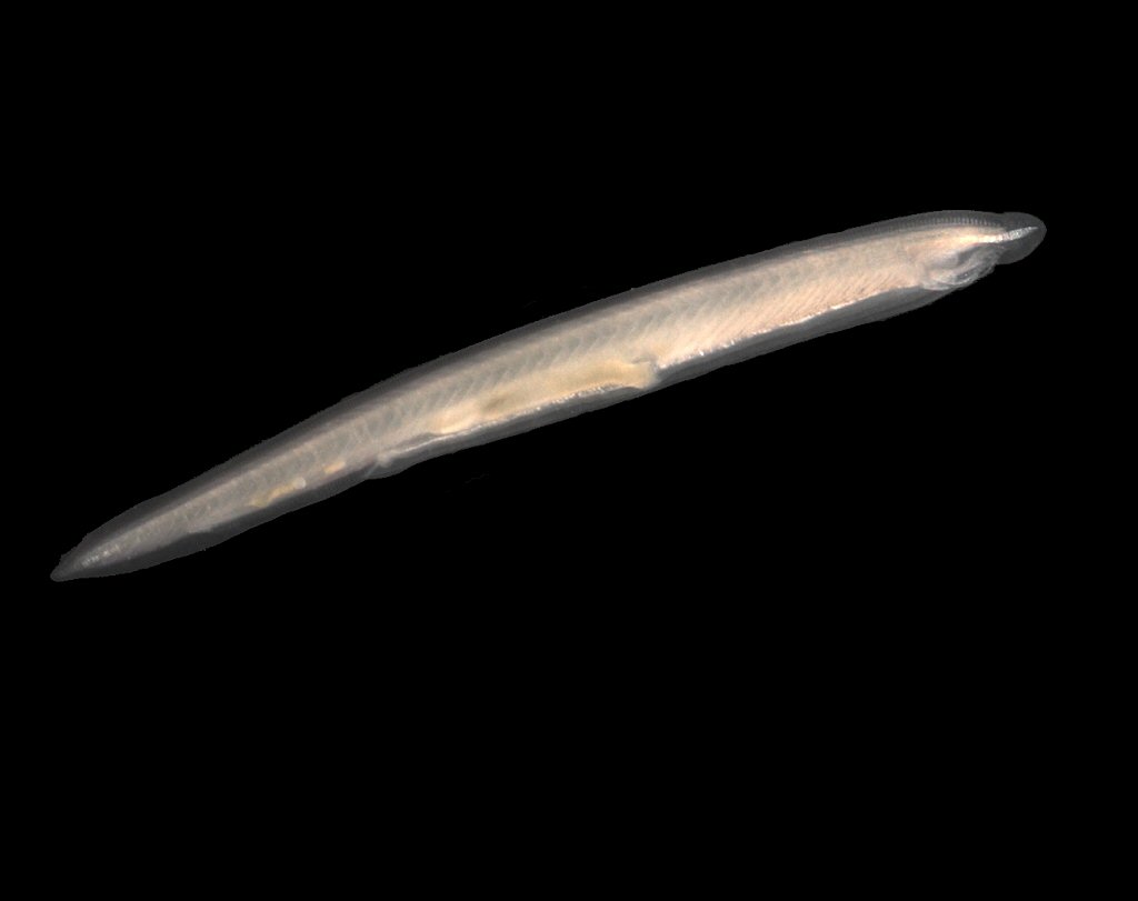
Perhaps smaller animals can skip the bone thing. Maybe a notable example are the lancelets, which look a bit like small fish. But they only go up to 8 cm.
Vertebrates minus tetrapods.
This paraphyletic subgroup is easy to form the "acquatic only" (fishes) vs "things that come out of water" (tetrapods). Though mudfishes make that distinction harder.
Which kind of makes sense, why would you want for limbs unless you are going to stay out of water!
Once Ciro joked in a twenty questions-like game that humans are animals.
But counting humans a fish would have been a stroke of genius.
Includes:
- amphibians
- amniotes, which includes:
- sauropsida: reptiles and birds, which really are reptiles
- mammals
The exact relationships between those clades is not very clear as there's a bunch of extinct species in the middle we are not sure exactly where they go exactly, some hypothesis are listed at: en.wikipedia.org/w/index.php?title=Tetrapod&oldid=1053601110#Temnospondyl_hypothesis_(TH)
But at least it seems rock solid that those three are actually clades.
Includes:
- sauropsida: reptiles and birds, which really are reptiles
- mammals, or if you want to include a bunch of extinct non-reptile mammal ancestors, synapsids.
Does not include amphibians. If you include them, you have the tetrapods.
This being a class is bullshit because it is not a clade, notably birds are not considered reptiles, but they are clearly in the clade.
TODO name: Wikipedia says "being with a fused arch" but what does that mean???
Good phylogenetic tree: en.wikipedia.org/w/index.php?title=Mammal&oldid=1052295685#Molecular_classification_of_placentals
Eggs are basal: they simply didn't evolve out of what other reptiles do. From which we conclude that milk came before eggs stopped.
So this is the most basal subclade of mammals.
Etymology: means "single hole" in Greek, because like other reptiles it has a single hole for shit, pee and fucking: the cloaca.
The name is completely random, "wild beast". Are platypuses not "wild beasts"? They have a freaking poison!!
They split up from the rest of the mammals after the monotremes.
Every other mammal has a placenta.
This baby in pouch thing just feels like a pre-placenta stage.
As of 2020, account for about 20% of the known mammal species!!! www.sciencefocus.com/nature/why-are-there-so-many-species-of-bat/ mentions some reasons:
- they can fly, so they can move out further
- their eating habits are highly specialized
When one specific species is implied, we will mean Mus musculus by default.
Exciting... sometimes cruel. But too exciting not to do:
Databases and projects:
- www.jax.org/research-and-faculty/resources/mouse-mutant-resource The Jackson Laboratory
Databases and projects:
- www.ncbi.nlm.nih.gov/pmc/articles/PMC2716027/ The Knockout Mouse Project (2004)
This is the level at which human and all extinct siblings lie, with no other extant species, all others were killed or fucked to death: Section "Interbreeding between archaic and modern humans".
Carnivores, ungulates, hedgehogs.
Tagged
- www.cell.com/cell-systems/fulltext/S2405-4712(16)30120-X
- www.cell.com/cell-systems/fulltext/S2405-4712(16)30151-X A Genome-Scale Database and Reconstruction of Caenorhabditis elegans Metabolism Gebauer, Juliane et al. Cell Systems , Volume 2 , Issue 5 , 312 - 322
Exactly 1033 somatic cells on male, 959 on hermaphrodite, every time, counted as of 2020. A beauty.
Exactly 131 commit apoptosis in the hermaphrodite.
www.wormatlas.org/celllineages.html contains the full lineage as some huge and impossible to view images. This image was taken directly from The embryonic cell lineage of the nematode Caenorhabditis elegans where it is split across many pages, it is a thing of beauty on the PDF.
www.wormatlas.org/celllistsulston.htm contains a non-hierarchical table with the cells and their names.
Tagged
A collection of closely related and curated C. elegans datasets.
This contains the C. elegans connectome.
The browseable thing is this massive interactive PDF: wormwiring.org/papers/Interactive-Diagram.pdf. It lists neurons from the C. elegans cell lineage using the standard cell names, and how they connect to each other. Some make a surprising ammount of connections.

Fantastic resouce that contains cross sections of C. elegans at various lengths of its body. Presumably frozen and cut with a Microtome and then scanned with electron microscopy.
Shame that there are so many parts missing.
It's the Visible Human Project, but for C. elegans!
TODO what does it contain. Does it have metabolic pathways?
This one has a 3D model of C. elegans containing all the cells, browsable on the browser at: browser.openworm.org/.

wormwideweb.org/
Browse freely moving whole-brain calcium imaging datasets
High level simulation only, no way to get from DNA to worm! :-) Includes:
- nervous system
- muscle system
3D body viewer at: browser.openworm.org/ TODO can you click on a cell to get its name?
OpenWorm Sibernetic demo by Mike Vella (2013)
Source. Sibernetic adds a fluid dynamics solver for brain-in-the-loop simulation of C. elegans.Formal name: "fungi".
Does not appear to refer to any one specific phylogenetic level, it usually refers to either:
- Saccharomyces cerevisiae, the model eukaryote unicellular organism
- two phila of the fungus kingdom
Size: 10 micrometers.
Genome:
Division time: 100 minutes.
Minimization project: en.wikipedia.org/wiki/Saccharomyces_cerevisiae#Synthetic_yeast_genome_project | syntheticyeast.org/
Formal name: "plantae".
This looks a lot like the beans that Brazilians venerate and can be easily found in the United Kingdom as of 2020.
The more exact type seems to be pinto bean, but this is close enough.
2021-03: same but 2.5 teaspons, seems to be the right ammount.
2021-02-10: attempt 3: 500g 1 hour 30 minutes no pressure, uncontrolled water. Salt with one chorizo: put 3 teaspoons, it was a bit too much, going to do 2 next time and see.
2020-12-14: attempt 3: 250g of beans, 1.5l of water, 30 minutes pressure.
2020-11-30: attempt 2: 275ml of dry beans, about 50% of 500g bag, putting 1650 ml (6x) of water on pressure cooker Still had to throw out some water.
Density dry raw: 216 g/250 ml = 432 g / 500 ml = 500 g / 580 ml = 864 g/L
500 g dry expands to in water after 12 hours: 1200 ml
Therefore 500 g dry = 864 / 2 L = 432 ml expands about 3x.
Therefore, to the maximum 2.5L of the cooker with 8x dry volume water from this recipe I can use:and so:which is about 227 / 580 = 40% of the 500 g bag.
2500 = volume expanded bean + volume water = 3 volume dry bean + 8 volume dry bean = 11 volume dry beanvolume dry bean = 2500/11 = 227mlAfter first try, I found that 8x volume of water is way, way too much. Going to try 6x next time.
This seems to be the "brown Brazilian bean" that many Brazilians eat every day.
Edit: after buying it, not 100% sure. This one felt smaller than what Ciro had in Brazil, borlotti beans might be closer. Pinto beans are smaller, and creamier, and have softer peel, possibly produced less natural gas.
2021-04: second try.
2021-03: did for first time, started with same procedure as borlotti beans 2021-03. Maybe 1h30 is too much. Outcome was still very good.